ABOUT US
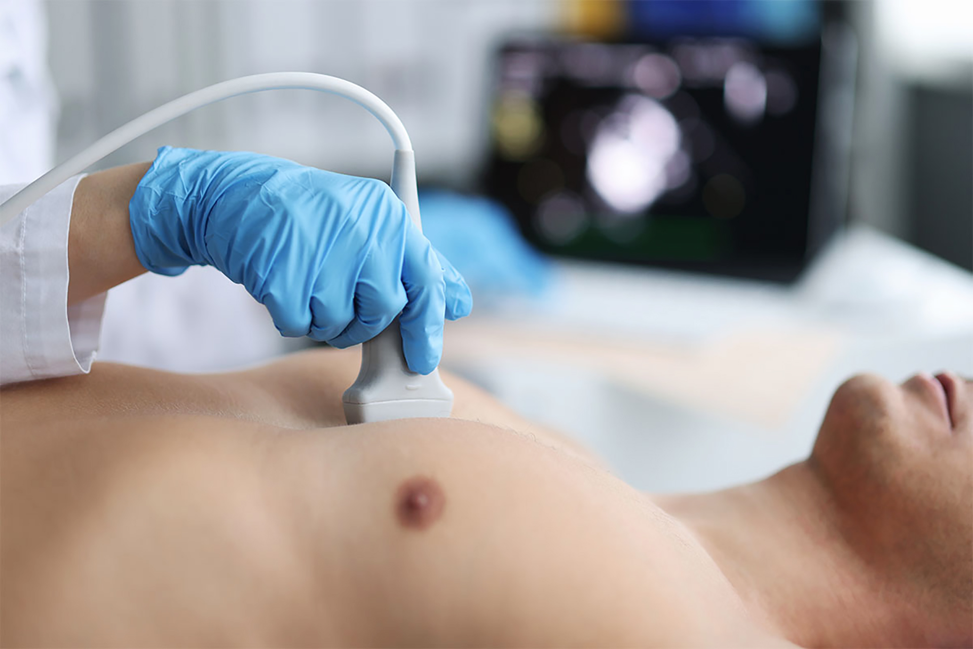
At Sonos Links Imaging, our mission is to deliver high-quality, patient-centered ultrasound services with a focus on compassion and care. We strive to ensure that every patient receives accurate diagnostic imaging in the most comfortable and convenient setting possible. We are one of the recognized industry leaders in providing affordable, high quality, and professional mobile ultrasound services focusing on the ultimate patient care.
OUR MISSION
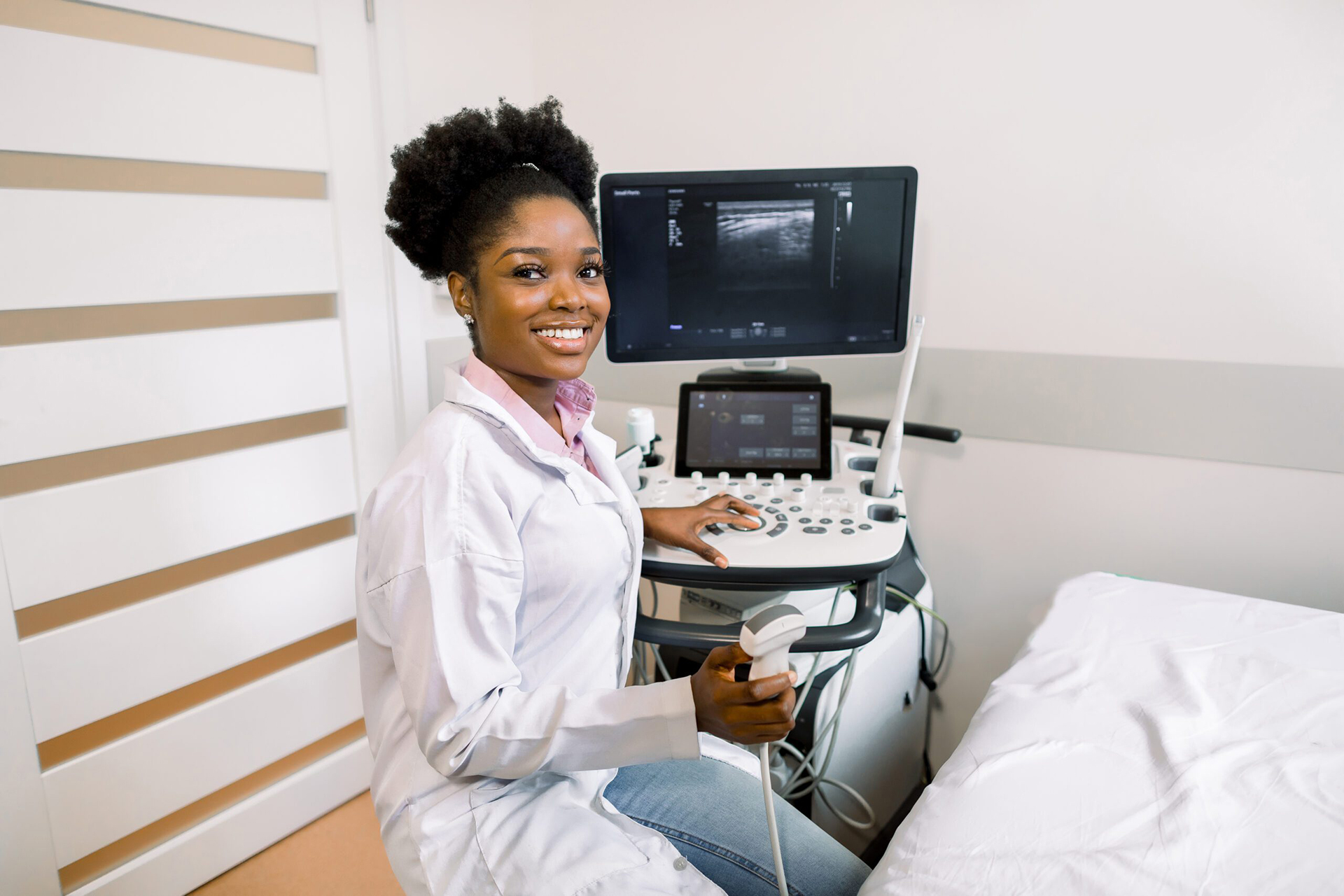
Bringing Compassionate Mobile Ultrasound Services to Southwest Florida.
Imagine receiving top-notch diagnostic care without the need to travel to a hospital or imaging center. For residents of assisted living facilities and skilled nursing homes, mobility can be a significant concern. Our mobile ultrasound services eliminate the stress and inconvenience of travel, providing essential diagnostic imaging right at your doorstep. This means less disruption to daily routines and a more comfortable experience for patients.